Male fruit. On whom and on what does the sex of the unborn child at conception depend: on chance, man or woman? Why exactly does a sperm with a Y chromosome fertilize an egg?
Pregnancy is a physiological process in which a new organism develops in the uterus, resulting from fertilization. Pregnancy lasts on average 40 weeks (10 obstetric months).
In the intrauterine development of a child, two periods are distinguished:
- Embryonic(up to 8 weeks of pregnancy inclusive). At this time, the embryo is called an embryo and acquires characteristic human features;
- Fetal(from 9 weeks until birth). At this time, the embryo is called a fetus.
The growth of a child, the formation of its organs and systems occurs naturally during various periods of intrauterine development, which is subject to the genetic code embedded in the germ cells and fixed in the process of human evolution.
Embryo development in the first obstetric month (1-4 weeks)
First week (days 1-7)
Pregnancy begins from the moment fertilization- fusion of a mature male cell (sperm) and a female egg. This process usually occurs in the ampullary section of the fallopian tube. After a few hours, the fertilized egg begins to divide exponentially and descends through the fallopian tube into the uterine cavity (this journey takes up to five days).
As a result of division turns out to be a multicellular organism, which is similar to a blackberry (in Latin “morus”), which is why the embryo at this stage is called Morula. Approximately on the 7th day, the morula penetrates the uterine wall (implantation). The villi of the outer cells of the embryo connect with the blood vessels of the uterus, and subsequently the placenta is formed from them. Other outer morula cells give rise to the development of the umbilical cord and membranes. Over time, various tissues and organs of the fetus will develop from the internal cells.
Information At the time of implantation, a woman may have slight bleeding from the genital tract. Such discharge is physiological and does not require treatment.
Second week (8-14 days)
The outer morula cells grow tightly into the lining of the uterus. In the embryo the formation of the umbilical cord and placenta begins, and neural tube, from which the fetal nervous system subsequently develops.
Third week (15-21 days)
The third week of pregnancy is a difficult and important period. At that time important organs and systems begin to form fetus: the rudiments of the respiratory, digestive, circulatory, nervous and excretory systems appear. At the site where the fetal head will soon appear, a wide plate is formed, which will give rise to the brain. On day 21, the baby's heart begins to beat.
Fourth week (22-28 days)
this week laying of fetal organs continues. The rudiments of the intestines, liver, kidneys and lungs are already present. The heart begins to work more intensely and pumps more and more blood through the circulatory system.
From the beginning of the fourth week in the embryo body folds appear, and appears vertebral primordium(chord).
Completed by day 25 neural tube formation.
By the end of the week (approximately 27-28 days) the muscular system and spine are formed, which divides the embryo into two symmetrical halves, both upper and lower limbs.
During this period it begins formation of pits on the head, which will later become the eyes of the fetus.
Development of the embryo in the second obstetric month (5-8 weeks)
Fifth week (29-35 days)
During this period the embryo weighs about 0.4 grams, length 1.5-2.5 mm.
The formation of the following organs and systems begins:
- Digestive system: liver and pancreas;
- Respiratory system: larynx, trachea, lungs;
- Circulatory system;
- Reproductive system: precursors of germ cells are formed;
- Sense organs: the formation of the eyes and inner ear continues;
- Nervous system: the formation of parts of the brain begins.
At that time a faint umbilical cord appears. The formation of limbs continues, the first rudiments of nails appear.
On the face the upper lip and nasal cavities are formed.
Sixth week (36-42 days)
Length embryo during this period is about 4-5 mm.
Starts in the sixth week formation of the placenta. At this stage, it is just beginning to function; blood circulation between it and the embryo has not yet been formed.
Ongoing formation of the brain and its parts. At the sixth week, when performing an encephalogram, it is already possible to record signals from the fetal brain.
Begins formation of facial muscles. The fetal eyes are already more pronounced and uncovered by eyelids that are just beginning to form.
During this period they begin upper limbs change: they lengthen and the rudiments of hands and fingers appear. The lower limbs still remain in their infancy.
Changes in important organs occur:
- Heart. The division into chambers is completed: ventricles and atria;
- Urinary system. Primary kidneys have formed, the development of the ureters begins;
- Digestive system. The formation of sections of the gastrointestinal tract begins: the stomach, small and large intestines. The liver and pancreas had practically completed their development by this period;
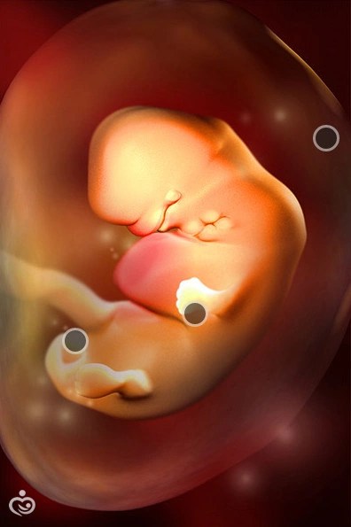
Seventh week (43-49 days)
The seventh week is significant in that it is finally The formation of the umbilical cord is completed and uteroplacental circulation is established. Now the breathing and nutrition of the fetus will be carried out due to blood circulation through the vessels of the umbilical cord and placenta.
The embryo is still bent in an arched manner; there is a small tail on the pelvic part of the body. The size of the head is at least half of the embryo. The length from the crown to the sacrum increases by the end of the week up to 13-15 mm.
Ongoing upper limb development. The fingers are visible quite clearly, but their separation from each other has not yet occurred. The child begins to perform spontaneous movements with his hands in response to stimuli.
Fine eyes are formed, already covered with eyelids, which protect them from drying out. The child can open his mouth.
The formation of the nasal fold and nose occurs, two paired elevations are formed on the sides of the head, from which they will begin to develop ears.
Intensive continues development of the brain and its parts.
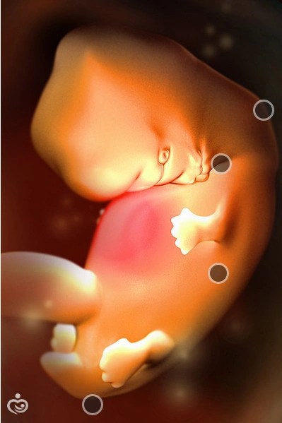
Eighth week (50-56 days)
The body of the embryo begins to straighten, length from the crown to the coccyx is 15 mm at the beginning of the week and 20-21 mm on day 56.
Ongoing formation of important organs and systems: digestive system, heart, lungs, brain, urinary system, reproductive system (boys develop testicles). The hearing organs are developing.
By the end of the eighth week the child's face becomes familiar to the person: the eyes are well defined, covered with eyelids, the nose, the ears, the formation of the lips is ending.
Intensive growth of the head, upper and lower horses is noted In particular, ossification of the long bones of the arms and legs and the skull develops. The fingers are clearly visible; there is no skin membrane between them.
Additionally At eight weeks the embryonic period of development ends and the fetal period begins. From this time on, the embryo is called a fetus.
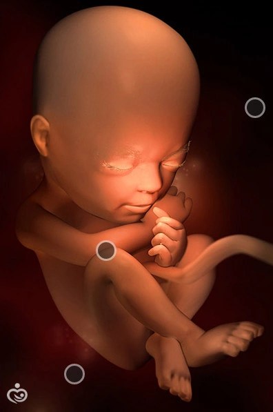
Fetal development in the third obstetric month (9-12 weeks)
Ninth week (57-63 days)
At the beginning of the ninth week coccygeal-parietal size fetus is about 22 mm, by the end of the week - 31 mm.
Happening improvement of blood vessels of the placenta, which improves uteroplacental blood flow.
The development of the musculoskeletal system continues. The process of ossification begins, the joints of the toes and hands are formed. The fetus begins to make active movements and can clench its fingers. The head is lowered, the chin is pressed tightly to the chest.
Changes occur in the cardiovascular system. The heart beats up to 150 times per minute and pumps blood through its blood vessels. The composition of blood is still very different from the blood of an adult: it consists only of red blood cells.
Ongoing further growth and development of the brain, cerebellar structures are formed.
The organs of the endocrine system are intensively developing, in particular, the adrenal glands, which produce important hormones.
Improves cartilage tissue: auricles, laryngeal cartilages, vocal cords are being formed.
Tenth week (64-70 days)
By the end of the tenth week fruit length from the coccyx to the crown is 35-40 mm.
Buttocks begin to develop, the previously existing tail disappears. The fetus is in the uterus in a fairly free position in a semi-bent state.
Nervous system development continues. Now the fetus performs not only chaotic movements, but also reflex ones in response to a stimulus. When accidentally touching the walls of the uterus, the child makes movements in response: turns his head, bends or straightens his arms and legs, and pushes to the side. The size of the fetus is still very small, and the woman cannot yet feel these movements.
The sucking reflex is formed, the child begins reflex movements with his lips.
The development of the diaphragm is completed, which will take an active part in breathing.
Eleventh week (71-77 days)
By the end of this week coccygeal-parietal size the fetus increases to 4-5 cm.
The fetal body remains disproportionate: small body, large head, long arms and short legs, bent at all joints and pressed to the stomach.
The placenta has already reached sufficient development and copes with its functions: ensures the supply of oxygen to the fetus and nutrients and removes carbon dioxide and metabolic products.
Further formation of the fetal eyes occurs: At this time, the iris develops, which will later determine the color of the eyes. The eyes are well developed, half-closed or wide open.
Twelfth week (78-84 days)
Coccygeal-parietal size fetus is 50-60 mm.
Goes clearly development of the genital organs according to the female or male type.
Happening further improvement of the digestive system. The intestines are elongated and arranged in loops, like those of an adult. Its periodic contractions begin - peristalsis. The fetus begins to make swallowing movements, swallowing amniotic fluid.
The development and improvement of the fetal nervous system continues. The brain is small in size, but exactly replicates all the structures of the adult brain. The cerebral hemispheres and other sections are well developed. Reflex movements are improved: the fetus can clench and unclench its fingers into a fist, grabs the thumb and actively sucks it.
In fetal blood Not only red blood cells are already present, but the production of white blood cells - leukocytes - also begins.
At this time the child single respiratory movements begin to be recorded. Before birth, the fetus cannot breathe, its lungs do not function, but it makes rhythmic movements of the chest, imitating breathing.
By the end of the week the fetus eyebrows and eyelashes appear, the neck is clearly visible.
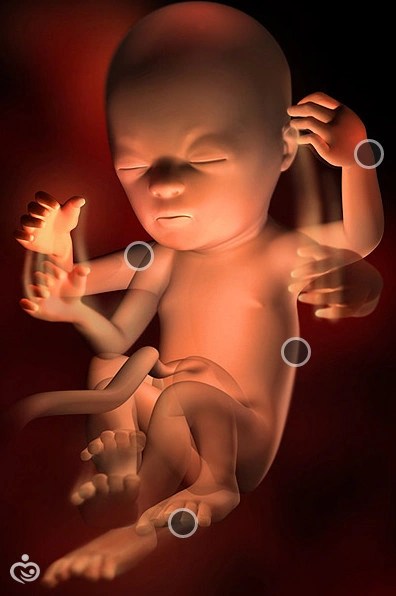
Fetal development in the fourth obstetric month (13-16 weeks)
Week 13 (85-91 days)
Coccygeal-parietal size by the end of the week is 70-75 mm. The proportions of the body begin to change: the upper and lower limbs and torso lengthen, the size of the head is no longer so large in relation to the body.
Improvement of the digestive and nervous systems continues. The embryos of baby teeth begin to appear under the upper and lower jaws.
The face is fully formed, the ears, nose and eyes are clearly visible (the eyelids are completely closed).
Week 14 (92-98 days)
Coccygeal-parietal size by the end of the fourteenth week it increases up to 8-9 cm. Body proportions continue to change to more familiar ones. The face has a well-defined forehead, nose, cheeks and chin. The first hair appears on the head (very thin and colorless). The surface of the body is covered with vellus hairs, which retain skin lubrication and thereby perform protective functions.
The musculoskeletal system of the fetus is improved. Bones become stronger. Motor activity increases: the fetus can turn over, bend, and make swimming movements.
Development of the kidneys, bladder and ureters is complete. The kidneys begin to secrete urine, which mixes with the amniotic fluid.
: pancreatic cells begin to work, producing insulin, and pituitary cells.
Changes in the genital organs appear. In boys, the prostate gland forms; in girls, the ovaries migrate into the pelvic cavity. At the fourteenth week, with a good sensitive ultrasound machine, it is already possible to determine the sex of the child.
Fifteenth week (99-105 days)
Coccygeal-parietal size of the fetus is about 10 cm, fruit weight – 70-75 grams. The head still remains quite large, but the growth of the arms, legs and torso begins to outstrip it.
The circulatory system is improved. In the fourth month, a child’s blood type and Rh factor can already be determined. Blood vessels (veins, arteries, capillaries) grow in length and their walls become stronger.
The production of original feces (meconium) begins. This is due to the ingestion of amniotic fluid, which enters the stomach, then into the intestines and fills it.
Fully formed fingers and toes, an individual design appears on them.
Sixteenth week (106-112 days)
The weight of the fetus increases to 100 grams, the coccygeal-parietal size - up to 12 cm.
By the end of the sixteenth week, the fetus is already fully formed, he has all the organs and systems. The kidneys are actively working, a small amount of urine is released into the amniotic fluid every hour.
The skin of the fetus is very thin, subcutaneous fatty tissue is practically absent, so blood vessels are visible through the skin. The skin appears bright red, covered with vellus hairs and grease. Eyebrows and eyelashes are well defined. Nails are formed, but they only cover the edge of the nail phalanx.
Facial muscles are formed, and the fetus begins to “grimace”: frowning of the eyebrows and a semblance of a smile are observed.
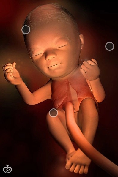
Fetal development in the fifth obstetric month (17-20 weeks)
Seventeenth week (days 113-119)
The weight of the fetus is 120-150 grams, the coccygeal-parietal size is 14-15 cm.
The skin remains very thin, but subcutaneous fatty tissue begins to develop under it. The development of baby teeth continues, which are covered with dentin. The embryos of permanent teeth begin to form under them.
There is a reaction to sound stimuli. From this week we can say for sure that the child began to hear. When strong sharp sounds appear, the fetus begins to actively move.
Fetal position changes. The head is raised and is in an almost vertical position. The arms are bent at the elbow joints, the fingers are clenched into a fist almost all the time. Periodically, the child begins to suck his thumb.
The heartbeat becomes clear. From now on, the doctor can listen to him using a stethoscope.
Eighteenth week (120-126 days)
The child's weight is about 200 grams, length - up to 20 cm.
The formation of sleep and wakefulness begins. Most of the time the fetus sleeps, movements stop during this time.
At this time, the woman may already begin to feel the baby moving, especially with repeated pregnancies. The first movements are felt as gentle jolts. A woman may feel more active movements when she is nervous or stressed, which affects the emotional state of the child. At this stage, the norm is about ten episodes of fetal movement per day.
Nineteenth week (127-133 days)
The child’s weight increases to 250-300 grams, body length – to 22-23 cm. The proportions of the body change: the head lags behind the body in growth, the arms and legs begin to lengthen.
Movements become more frequent and noticeable. They can be felt not only by the woman herself, but also by other people by placing their hand on their stomach. Primigravidas at this time can only begin to feel movements.
The endocrine system is improved: the pancreas, pituitary gland, adrenal glands, gonads, thyroid and parathyroid glands are actively functioning.
Blood composition has changed: In addition to erythrocytes and leukocytes, the blood contains monocytes and lymphocytes. The spleen begins to take part in hematopoiesis.
Twentieth week (134-140 days)
Body length increases to 23-25 cm, weight – up to 340 grams.
The fetal skin is still thin, covered with protective lubricant and vellus hairs, which can persist until childbirth. Subcutaneous fatty tissue develops intensively.
Well formed eyes, at twenty weeks the blink reflex begins to appear.
Improved movement coordination: The child confidently brings his finger to his mouth and begins to suck it. Facial expressions are pronounced: the fetus may close its eyes, smile, or frown.
This week all women are already feeling movements., regardless of the number of pregnancies. Movement activity varies throughout the day. When stimuli appear (loud sounds, stuffy rooms), the child begins to move very violently and actively.
Fetal development in the sixth obstetric month (21-24 weeks)
Twenty-first week (days 141-147)
Body weight grows to 380 grams, fetal length – up to 27 cm.
The layer of subcutaneous tissue increases. The skin of the fetus is wrinkled, with many folds.
Fetal movements become more active and tangible. The fetus moves freely in the uterine cavity: it lies head down or buttocks, across the uterus. Can pull on the umbilical cord, push off the walls of the uterus with hands and feet.
Changes in sleep and wakefulness patterns. Now the fetus spends less time sleeping (16-20 hours).
Twenty-second week (148-154 days)
At week 22, the size of the fetus increases to 28 cm, weight - up to 450-500 grams. The size of the head becomes proportional to the body and limbs. The legs are bent almost all the time.
The fetal spine is fully formed: It has all the vertebrae, ligaments and joints. The process of strengthening bones continues.
Improves the fetal nervous system: The brain already contains all the nerve cells (neurons) and has a mass of about 100 grams. The child begins to take an interest in his body: he feels his face, arms, legs, tilts his head, brings his fingers to his mouth.
Heart size increases significantly, the functionality of the cardiovascular system is improved.
Twenty-third week (155-161 days)
The length of the fetal body is 28-30 cm, weight is about 500 grams. Pigment begins to be synthesized in the skin, resulting in the skin becoming bright red. The subcutaneous fatty tissue is still quite thin, as a result the child looks very thin and wrinkled. The lubricant covers the entire skin and is more abundant in the folds of the body (elbow, axillary, inguinal, etc. folds).
Development of internal genital organs continues: in boys - the scrotum, in girls - the ovaries.
Respiratory frequency increases up to 50-60 times per minute.
The swallowing reflex is still well developed: the child constantly swallows amniotic fluid with particles of protective skin lubricant. The liquid part of the amniotic fluid is absorbed into the blood, leaving a thick green-black substance (meconium) in the intestines. Normally, the bowel should not have a bowel movement until the baby is born. Sometimes swallowing water causes hiccups in the fetus; a woman can feel it in the form of rhythmic movements for several minutes.
Twenty-fourth week (162-168 days)
By the end of this week, the weight of the fetus increases to 600 grams, body length to 30-32 cm.
The movements are becoming stronger and clearer. The fetus takes up almost all the space in the uterus, but can still change position and turn over. Muscles grow rapidly.
By the end of the sixth month, the child has well-developed sense organs. Vision begins to function. If a bright light hits a woman’s belly, the fetus begins to turn away and closes her eyelids tightly. Hearing is well developed. The fetus determines pleasant and unpleasant sounds for itself and reacts to them differently. When hearing pleasant sounds, the child behaves calmly, his movements become calm and measured. When unpleasant sounds occur, it begins to freeze or, conversely, moves very actively.
An emotional connection is established between mother and child. If a woman experiences negative emotions (fear, anxiety, melancholy), the child begins to experience similar feelings.
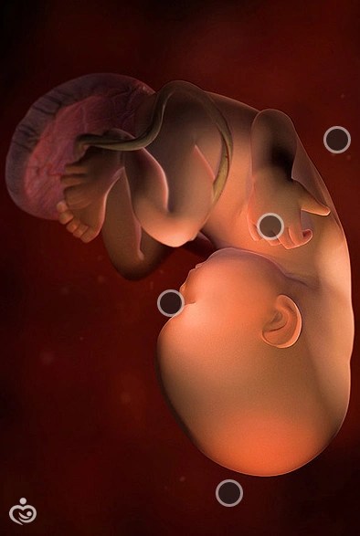
Fetal development in the seventh obstetric month (25-28 weeks)
Twenty-fifth week (169-175 days)
The length of the fetus is 30-34 cm, body weight increases to 650-700 grams. The skin becomes elastic, the number and severity of folds decreases due to the accumulation of subcutaneous fatty tissue. The skin remains thin with a large number of capillaries, giving it a red color.
The face has a familiar appearance to a person: eyes, eyelids, eyebrows, eyelashes, cheeks, ears are well defined. The cartilage of the ears remains thin and soft, their bends and curls are not fully formed.
Bone marrow develops intensively, which takes on the main role in hematopoiesis. The strengthening of the fetal bones continues.
Important processes occur in lung maturation: small elements of lung tissue (alveoli) are formed. Before the baby is born, they are without air and resemble deflated balloons, which straighten out only after the first cry of the newborn. From week 25, the alveoli begin to produce a special substance (surfactant) necessary to maintain their shape.
Twenty-sixth week (176-182 days)
The length of the fruit is about 35 cm, the weight increases to 750-760 grams. The growth of muscle tissue and subcutaneous fat continues. Bones are strengthened and permanent teeth continue to develop.
The formation of the genital organs continues. In boys, the testicles begin to descend into the scrotum (the process lasts 3-4 weeks). In girls, the formation of the external genitalia and vagina is completed.
Improved sense organs. The child develops a sense of smell (smell).
Twenty-seventh week (183-189 days)
Weight increases to 850 grams, body length - up to 37 cm.
The organs of the endocrine system are actively functioning, in particular the pancreas, pituitary gland and thyroid gland.
The fruit is quite active, makes freely various movements inside the uterus.
From the twenty-seventh week in the child individual metabolism begins to form.
Twenty-eighth week (190-196 days)
The child’s weight increases to 950 grams, body length – 38 cm.
By this age the fetus becomes practically viable. In the absence of organ pathology, a child with good care and treatment can survive.
Subcutaneous fat continues to accumulate. The skin is still red in color, the vellus hair begins to gradually fall out, remaining only on the back and shoulders. Eyebrows, eyelashes, and hair on the head become darker. The child begins to open his eyes frequently. The cartilage of the nose and ears remains soft. The nails do not yet reach the edge of the nail phalanx.
This week starts more one of the cerebral hemispheres is actively functioning. If the right hemisphere becomes active, then the child becomes left-handed; if the left hemisphere becomes active, then right-handedness develops.
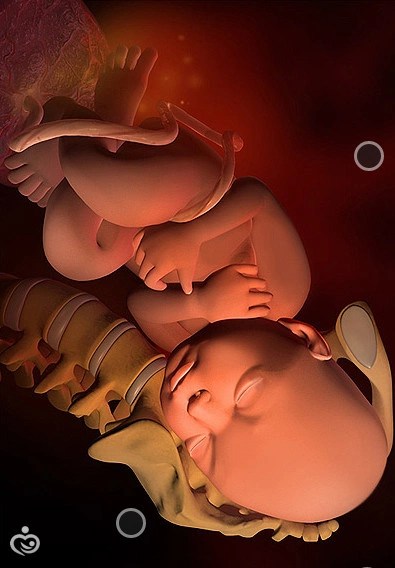
Fetal development in the eighth month (29-32 weeks)
Twenty-ninth week (197-203 days)
The weight of the fetus is about 1200 grams, the height increases to 39 cm.
The baby has already grown enough and takes up almost all the space in the uterus. Movements become less chaotic. The movements manifest themselves in the form of periodic kicks with the legs and arms. The fetus begins to take a definite position in the uterus: head or buttocks down.
All organ systems continue to improve. The kidneys already secrete up to 500 ml of urine per day. The load on the cardiovascular system increases. The blood circulation of the fetus is still significantly different from the blood circulation of the newborn.
Thirtieth week (204-210 days)
Body weight increases to 1300-1350 grams, height remains approximately the same - about 38-39 cm.
Subcutaneous fat tissue constantly accumulates, skin folds straighten out. The child adapts to the lack of space and takes a certain position: curls up, arms and legs crossed. The skin still has a bright color, the amount of grease and vellus hair decreases.
Alveolar development and surfactant production continues. The lungs prepare for the birth of the baby and the start of breathing.
Brain development continues brain, the number of convolutions and the area of the cortex increases.
Thirty-first week (211-217 days)
The child's weight is about 1500-1700 grams, height increases to 40 cm.
Your child's sleep and wake patterns change. Sleep still takes a long time, during which time there is no motor activity of the fetus. While awake, the child actively moves and pushes.
Fully formed eyes. During sleep, the child closes his eyes, while awake, the eyes are open, and the child blinks periodically. The color of the iris is the same for all children (blue), then after birth it begins to change. The fetus reacts to bright light by constricting or dilating the pupil.
Brain size increases. Now its volume is about 25% of the volume of the adult brain.
Thirty-second week (218-224 days)
The child's height is about 42 cm, weight - 1700-1800 grams.
Accumulation of subcutaneous fat continues, due to which the skin becomes lighter, there are practically no folds left on it.
Are improving internal organs : organs of the endocrine system intensively secrete hormones, surfactant accumulates in the lungs.
The fetus produces a special hormone, which promotes the formation of estrogen in the mother’s body, as a result, the mammary glands begin to prepare for milk production.
Fetal development in the ninth month (33-36 weeks)
Thirty-third week (225-231 days)
The weight of the fetus increases to 1900-2000 grams, the height is about 43-44 cm.
Skin becomes increasingly lighter and smoother, the layer of fatty tissue increases. The vellus hair is increasingly wiped off, and the layer of protective lubricant, on the contrary, increases. Nails grow to the edge of the nail phalanx.
The baby becomes increasingly cramped in the uterine cavity, so his movements become more rare, but strong. The position of the fetus is fixed (head or buttocks down), the likelihood that the child will turn over after this period is extremely small.
The functioning of internal organs is becoming more and more improved: the mass of the heart increases, the formation of the alveoli is almost complete, the tone of the blood vessels increases, the brain is fully formed.
Thirty-fourth week (232-238 days)
The child's weight ranges from 2000 to 2500 grams, height is about 44-45 cm.
The baby now occupies a stable position in the uterus. The bones of the skull are soft and mobile thanks to the fontanelles, which can close only a few months after birth.
Head hair grows rapidly and take on a certain color. However, hair color may change after childbirth.
Intensive strengthening of bones is noted, in connection with this, the fetus begins to take calcium from the mother’s body (the woman may notice the appearance of cramps at this time).
The child constantly swallows amniotic fluid, thereby stimulating the gastrointestinal tract and kidney function, which secrete at least 600 ml of clear urine per day.
Thirty-fifth week (239-245 days)
Every day the child gains 25-35 grams. Weight during this period can vary greatly and by the end of the week it is 2200-2700 grams. Height increases to 46 cm.
All internal organs of the child continue to improve, preparing the body for the upcoming extrauterine existence.
Fatty tissue is intensively deposited, the child becomes more well-fed. The amount of vellus hair is greatly reduced. The nails have already reached the tips of the nail phalanges.
A sufficient amount of meconium has already accumulated in the fetal intestines, which normally should go away 6-7 hours after birth.
Thirty-sixth week (246-252 days)
The weight of a child varies greatly and can range from 2000 to 3000 grams, height - within 46-48 cm
The fetus already has well-developed subcutaneous fatty tissue, skin color becomes lighter, wrinkles and folds disappear completely.
The baby occupies a certain position in the uterus: more often he lies upside down (less often, with his legs or buttocks, in some cases, transversely), his head is bent, his chin is pressed to his chest, his arms and legs are pressed to his body.
Skull bones, unlike other bones, remain soft, with cracks (fontanelles), which will allow the baby's head to be more pliable when passing through the birth canal.
All organs and systems are fully developed for the existence of a child outside the womb.
Fetal development in the tenth obstetric month
Thirty-seventh week (254-259 days)
The child's height increases to 48-49 cm, weight can fluctuate significantly. The skin has become lighter and thicker, the fat layer increases every day by 14-15 grams per day.
Cartilages of the nose and ears become denser and more elastic.
Fully lungs are formed and mature, the alveoli contain the necessary amount of surfactant for the newborn to breathe.
The digestive system has matured: Contractions occur in the stomach and intestines to push food through (peristalsis).
Thirty-eighth week (260-266 days)
A child's weight and height vary greatly.
The fetus is fully mature and ready to be born. Externally, the child looks like a full-term newborn. The skin is light, the fatty tissue is sufficiently developed, and vellus hair is practically absent.
Thirty-ninth week (267-273 days)
Typically two weeks before birth the fruit begins to descend, pressing against the pelvic bones. The child has already reached full maturity. The placenta begins to gradually age and its metabolic processes deteriorate.
The weight of the fetus increases significantly (30-35 grams per day). The proportions of the body change completely: the chest and shoulder girdle are well developed, the belly is round, and the limbs are long.
Well developed sense organs: the child catches all sounds, sees bright colors, can focus his vision, and taste buds are developed.
Fortieth week (274-280 days)
All indicators of fetal development correspond to new to the awaited one. The baby is completely ready for birth. The weight can vary significantly: from 250 to 4000 and above grams.
The uterus begins to periodically contract(), which is manifested by aching pain in the lower abdomen. The cervix opens slightly, and the fetal head is pressed closer to the pelvic cavity.
The skull bones are still soft and pliable, which allows the baby’s head to change shape and pass the birth canal more easily.
Fetal development by week of pregnancy - Video
Pregnancy is the process in which a baby develops from two tiny parent cells. The development of the fetus by week of pregnancy is a fascinating story about what exactly happens in each week of pregnancy, how the weight and height of the fetus changes, what sensations arise in the mother as the pregnancy lengthens. In the article we will tell you about what interests every expectant mother: when the baby begins to hear her speech, when and how the weight of the fetus changes, when you can take a photo of the fetus with an ultrasound, what causes the mother’s feelings during pregnancy, and much more.
First and second weeks of pregnancy: baby? Which child?
At the moment the embryo appears, the pregnancy period is already 2 weeks. Why? Let's decide from what we will calculate the period. There are concepts of embryonic and obstetric term. The embryonic period of pregnancy is the true period from the moment of conception. Obstetric period - from the first day of the last menstruation. The obstetric period is on average 2 weeks longer than the embryonic period. During an ultrasound, the pregnant woman’s chart and sick leave will always indicate the obstetric period according to the date of the last menstruation. But from the third week of pregnancy, the development of the fetus actually begins. Below you will find a description of each week of pregnancy: how the fetus develops, what happens to the uterus, how the expectant mother’s sensations change.
3rd week of pregnancy: parents meeting

At the end of the second and beginning of the third week (on average on the 14th day of the cycle), ovulation occurs. At this moment, the woman’s egg leaves the ovary into the fallopian tube and where it meets the sperm in the next 24 hours. Of the 75-900 million sperm that enter the vagina, less than a thousand reach the cervical canal. And only one will penetrate the egg.
The sperm and egg carry half the set of chromosomes of the future person. As a result of their fusion, the first cell of a new organism with a complete set of chromosomes is formed - a zygote. Chromosomes determine the baby's gender, eye color, and even character. The zygote begins to divide and move towards the uterine cavity. The journey to the uterus will take approximately 5 days, by which time the embryo will consist of approximately 100 cells. The next stage is implantation - the introduction of the embryo into the wall of the uterus.
4th week of pregnancy

The ball of cells is officially called an embryo. The size of the fruit at this stage is like a poppy seed, approximately 1.5 mm.
At the end of this week, the expectant mother notices that the expected menstruation does not begin. At this time, a woman may feel drowsiness, weakness, increased sensitivity of the mammary glands, and mood swings. A pregnancy test shows a positive result. The test detects the hCG hormone, which begins to be produced after implantation.
The embryonic period lasts up to 12 weeks. The axial organs and tissues of the baby are formed. A yolk sac with a supply of nutrients and an amniotic sac are formed; from these extra-embryonic organs, the fetal membranes and chorion - the future placenta - subsequently develop. Below we will look at what happens in the embryonic period every week, how the height and weight of the fetus changes and what sensations await the woman.
5th week of pregnancy

The embryo consists of three layers - the outer ectoderm, from which the ears, eyes, inner ear, and connective tissue will be formed; endoderm, from which the intestines, bladder and lungs will develop; and mesoderm - the basis for the cardiovascular system, bones, muscles, kidneys, and reproductive organs.
The anterior and posterior poles of the embryo are determined - the future head and legs. The body of the embryo is laid along the axis of symmetry - the chord. All organs will be symmetrical. Some are paired, for example, kidneys. Others grow from symmetrical buds, such as the heart and liver.
At the 5th week of pregnancy, with a hCG level of 500-1000 IU/l, a fertilized egg can be determined to be 2 mm in size, which is the size of a sesame seed. Each woman experiences this period differently, but most experience nausea, drowsiness, and odor intolerance - signs of toxicosis.
6th week of pregnancy
Now the baby is no larger than a lentil, at the beginning of the week 3 mm, and by the end - 6-7 mm. The embryo is somewhat similar to a fish and still bears little resemblance to a person. The rudiments of arms and legs appear. When the hands appear, the legs will still be in the form of rudiments. The cerebral hemispheres are formed. The small heart is pulsating and is dividing into sections.
The future placenta is formed from chorionic villi, vessels actively grow through which blood is exchanged, and accordingly everything necessary for the unborn child between mother and baby.
At this time, toxicosis may intensify, and severe weakness and vomiting may appear. Drinking enough fluids is important during these weeks of pregnancy.
7th week of pregnancy
The embryo is approximately the size of a blueberry, height 8-11 mm, weight up to 1 g. Hints of the future nose, eyes, ears and mouth appear. There is a fantastic rate of brain growth - 100,000 cells per minute! Interdigital spaces have already appeared on the handles, but the fingers are not yet separated. The umbilical cord and the uteroplacental circulatory system are formed: the baby’s breathing and nutrition comes from the mother’s blood.
It is during this period that many expectant mothers often come for their first ultrasound scan during pregnancy. At 7-8 weeks, the CTE (coccygeal-parietal size) is 10-15 mm. An ultrasound detects a heartbeat with a frequency of 100 to 190 beats per minute, which is significantly higher than that of an adult. At this time, the first photo of the gallery of fetal development is taken week by week. Without a doctor's instructions, you won't know where to look. It will be clearer later, especially on a 3D ultrasound.
While the mother has not yet noticed an enlarged abdomen, the gynecologist can already tell about an enlarged uterus. The woman experiences increased urination, which is associated with an increase in the volume of fluid in the body.
8th week of pregnancy

The baby is the size of a bean, from 15 to 40 mm, and weighs approximately 5 grams. Over the past two weeks it has grown 4 times! The contours of the face continue to develop, they become more graceful, the upper lip and tip of the nose stand out, and the formation of the eyelids begins.
At the 8th week of pregnancy, ossification of bones begins - arms, legs, skull. The structuring of the gastrointestinal tract, heart, kidneys, and bladder is completed.
Around 7-8 weeks of pregnancy, the baby begins to move, but the mother will not feel these movements in the coming months. Mom’s condition remains virtually unchanged. It may become easier due to adaptation to the condition and awareness of your new role.
9th week of pregnancy

The little man is only the size of a grape - its length is 35-45 mm, and its weight reaches 10 grams. The formation of the reproductive system occurs, and the adrenal glands are already producing hormones, including adrenaline.
The brain is rapidly developing, including the cerebellum, which is responsible for coordinating movements. Movements become more controlled. The digestive system is actively developing. The liver begins to produce new blood cells. The head occupies half the entire length of the body. Tiny fingers are getting longer.
The amount of circulating fetal DNA in the mother's blood is sufficient to perform a non-invasive prenatal test.
The mother still has signs of toxicosis. Usually at this time she turns to a gynecologist to register.
10th week of pregnancy

Do you know this fruit - kumquat? This is approximately the size of the baby now. This week it will officially be called a fetus, but for now we call it an embryo. This period is considered the end of the first critical period. Now the dangerous effect of drugs leading to developmental defects is not so significant.
A lot of events are happening these days. The webbing between the fingers disappears and the fingers separate. The bones harden. The kidneys begin to work, performing their main function - producing urine. The brain produces 250,000 neurons every minute. A diaphragm is formed between the abdominal and thoracic cavities.
My mother is experiencing symptoms of toxicosis. Due to changes in nutrition, metabolism, muscle tone and hormonal fluctuations, your figure and body movements may change. The uterus is the size of a grapefruit, but the pregnancy is not yet noticeable to others.
11th week of pregnancy

From 11 to 13 weeks, the baby undergoes a serious medical examination - ultrasound screening. The thickness of the collar space and nasal bones are determined, a study of blood vessels is carried out, and gross changes in the structure of the body are excluded. They examine the internal organs, facial structure, brain, arms and legs, and spine. Your baby is only the size of a fig, and the doctor describes the anatomy of the fetus in such detail! The head is still large relative to the body, but the proportions continue to change: the head is large, the body is small, the upper limbs are long, and the lower ones are short and bent at the knees. The rudiments of nails and teeth appear.
With the results of the ultrasound, the mother is given a biochemical blood test to check for chromosomal abnormalities and the risk of developing pregnancy complications.
The symptoms of toxicosis are replaced by new sensations: heartburn, bloating, and there may be constipation. A woman should pay more attention to her diet and fluid intake.
12th week of pregnancy
Your baby is about the size of a lime. Until 11-12 weeks, there are no reliable ultrasound differences between boys and girls. The probability of correctly determining the sex of the fetus is already above 50%. The weight of the fruit is about 20 grams, and the length is about 9 cm.
At this stage, the baby begins to actively move his arms and legs, hands, and fingers. Due to active growth, the intestines no longer fit in the tummy and begin to fold into loops. During this period, the intestines are trained: amniotic fluid passes through it, which is swallowed by the fetus. White blood cells appear in the blood - leukocytes, which have the function of protecting against infections.
Mom's weight gain About the norms of 12 weeks of development and ultrasound of pregnancy - about 1-2 kg. Doctors recommend doing gymnastics for pregnant women, and swimming is recommended.
13th week of pregnancy

A pea pod - this is how you can describe the size of a baby in everyday measurements. Or 7-10 cm, 20-30 grams. From the 13th week the second trimester of pregnancy begins. All the main organs and systems have already been formed; the rest of the time before birth, the organs will grow and develop.
The face becomes more and more human-like. The ears move closer and closer to their place from the neck, and the eyes from the side to the center of the face. The first hairs appear. 20 baby teeth have been formed.
The head is still disproportionately large, but the body will now grow faster. The hands continue to grow, the baby can already reach his face. Often, during an ultrasound, doctors show parents how the baby puts his finger in his mouth.
During this period, the shape of the abdomen changes, and the previous clothes become tight. Those around you may notice a woman’s new emotional mood; she becomes calmer and more relaxed.
14th week of pregnancy

At week 14, the fetus grows to 13 cm and 45 grams. In boys, the prostate forms, and in girls, the ovaries descend into the pelvis. The palate is already fully formed, active reflex sucking begins. The baby imitates breathing movements in order to effectively take the first breath after birth.
The formed pancreas begins to produce the most important hormone of carbohydrate metabolism - insulin. And in the depths of the brain, the pituitary gland begins to work - the head of all organs of the endocrine system, it is he who subsequently controls all the glands of the body.
The uterus is located 10-15 cm above the pubis; the woman herself can feel its upper part. It is recommended to use special cosmetics for the skin of the abdomen.
15th week of pregnancy

The fruit is the size of an apple and weighs about 70 grams. The whole baby is covered with small vellus hair - they are on the back, shoulders, ears, and forehead. These hairs help retain heat. Then, when the baby gains enough fatty tissue, the hairs will fall off. The child makes various grimaces, winces, frowns, squints, but this does not reflect his mood at all. He changes his position all the time, actively moving. But the baby is still too small and does not hit the walls of the uterus. A unique skin pattern appears on the fingertips and special proteins appear on the red blood cells that determine the blood type.
Mom may develop pigmentation on her stomach.
16th week of pregnancy

The baby is the size of an avocado. Skeletal bones become harder, but flexible enough for the baby to pass through the birth canal. The umbilical cord contains one vein and two arteries, surrounded by a gelatinous substance that protects the vessels from pinching and makes the umbilical cord slippery for movement. Girls these days are forming sex cells - your future grandchildren.
Weight gain during this week of pregnancy is 2-3 kg.
17th week of pregnancy

The baby's size is 12-13 cm and weighs up to 150 g, the size of a turnip. The arms and legs are commensurate with the size of the body and head. Fat begins to be deposited under the skin and sweat glands develop. The placenta provides the baby with vitamins, minerals, proteins, fats and oxygen while removing waste products.
Due to an increase in the volume of circulating blood, the mother may experience a rapid heartbeat. In this case, bring this to the attention of your doctor to see if everything is okay.
18th week of pregnancy
Your child is the size of a bell pepper and weighs 250 grams and is ready to communicate. Yes, now the baby can hear, and a loud sound can scare him. He gets used to his parents' voice, and will soon be able to recognize it from other sounds.
The fetal endocrine system is actively developing and functioning. There are so many “baby” hormones that the baby can even supply the mother’s body.
This week, mom may feel fetal movements for the first time. As long as they are mild and infrequent, don't worry if you don't hear your baby too often.
19th week of pregnancy

The height of the fetus is 25 cm, and the weight is already 250-300 grams.
The cheese-like lubricant coats your baby's skin and helps regulate body temperature. The formation of molars occurs; they are located under the rudiments of milk teeth. The head does not grow as quickly, but the limbs and body continue to grow, so the baby becomes more symmetrical.
The uterus is located 1-2 cm below the navel. Due to its intensive growth, painful sensations associated with stretching of the uterine ligaments may occur.
20th week of pregnancy

A happy child weighing 240 grams. At this stage, he is especially good at flexing and extending his arms and legs. He is becoming more and more like his parents.
Week 20 is the equator of pregnancy. The growing uterus compresses the internal organs, so the mother experiences shortness of breath and frequent urination.
During these weeks, the mother attends the next scheduled ultrasound and Doppler measurements are performed. This is a good time for a video ultrasound and regular photos of the heir.
21st week of pregnancy

The height of the fetus is 25 cm, and the weight is 400 g. Most of the nutrients come from the placenta. If amniotic fluid is swallowed, the stomach is already equipped to digest it and receive nutrients. The baby begins to feel the taste.
The mother gains more weight as the baby grows rapidly.
22nd week of pregnancy

By the end of the week the baby will be about 500 grams. The skin is no longer transparent, but remains red and wrinkled and covered with grease. Nerve endings mature and the baby becomes sensitive to touch. From 21 to 25 weeks, the brain increases 5 times - from 20 to 100 grams!
23rd week of pregnancy
Billions of brain cells will develop over the next few weeks. Their job is to monitor all of your baby's movements, senses, and basic life functions such as breathing.
The lungs begin to produce a substance that allows the lungs to inflate and fill with air after birth, and the fetus begins to “breathe.” The frequency of respiratory movements is 50-60 per minute.
The height of the uterine fundus is 4 cm above the navel. The uterus grows, which can cause discomfort in the spine and joints, so a special bandage may be required.
24th week of pregnancy
The baby is still small, his weight is 600 g, and his height is about 33 cm. The child actively reacts when addressed to him. The inner ear is already fully formed (the vestibular apparatus), he began to understand where is up and where is down, movements in the uterine cavity become more meaningful.
Mom gains about 500 grams per week. Swelling of the feet may occur, so it is important to choose comfortable shoes and rest your feet.
25th week of pregnancy

The height of the fetus is 30-32 cm, weight is 750 grams. Meconium forms in the large intestine - the baby's first stool, which will be completely passed within a few days after birth. The osteoarticular system is actively developing, bones are strengthening.
The mother may experience signs of anemia (anemia) developing due to iron deficiency. Fatigue, pallor, fatigue and tachycardia are reasons to consult a therapist and take blood tests for anemia.
26th week of pregnancy

Height 34 cm, weight 900 grams.
The lungs are actively developing, they are filled with a special substance that will not allow the lungs to stick together after the first breath.
The child has clearly defined periods of sleep and wakefulness. Mom feels his activity by movement in her stomach. If you are lucky, your sleep and activity periods will coincide with your baby's.
27th week of pregnancy

The fetal body weight is already about a kilogram, and its height is 34 cm. Growth hormone begins to be produced in the pituitary gland. And the thyroid gland contains hormones that regulate metabolism.
Due to involuntary contractions of the diaphragm, the mother may feel hiccup-like movements of the baby. In adults, such movements are accompanied by the closure of the vocal cords, which is why the characteristic “hiccup” sound occurs, but in a baby before birth, this space is filled with liquid, so this “hiccup” is silent.
You may experience new sensations in your legs—tingling, pins and needles, or even cramps. This is a reason to consult a doctor for further examination and treatment.
28th week of pregnancy

Now your baby is beginning to close and open his eyes, which until this moment were not completely closed. The iris of the eyes acquired color due to pigment, although this coloring is not final. Children's eye color may change until they are one year old.
At week 28, in case of multiple pregnancy, the mother receives a “sick leave”. The weight gain by this time is 7-9 kg. At this time, Rh-negative mothers are given immunoglobulin.
29th week of pregnancy

The child is 36-37 cm long, weighs approximately 1300 grams and is becoming stronger and more active. We can say that he shows character. A child reacts differently to different foods, sounds, and light.
A woman suffers from heartburn and heaviness after eating. There may be frequent urination or even false urges.
30th pregnancy

In the coming weeks, your baby will actively gain weight. Adipose tissue will perform the function of thermoregulation after birth, provide energy, and protect organs. The baby's movements will become less active, which is associated with an increase in his size. But if you do not feel any shocks as usual, be sure to tell your doctor. Pregnant women may feel breast swelling and notice the release of colostrum.
At this time, a certificate of incapacity for work is issued for a singleton pregnancy.
From 28-30 weeks of pregnancy, regular CTG (cardiotocography) begins to assess the condition of the fetus. CTG evaluates fetal heartbeat, uterine tone and motor activity.
31st week of pregnancy

Before birth, the little man will be in the fetal position, because otherwise he will no longer fit in the uterine cavity, his weight is 1600 g, and his height is already 40 cm.
These days, an important event occurs in male fetuses - the testicles on the way to the scrotum. In girls, the clitoris is almost formed.
Weekly weight gain is 300-400 grams. During these periods, swelling may appear and blood pressure may increase, which can be a symptom of a serious complication - preeclampsia. Therefore, the expectant mother should be as attentive as possible to any changes in well-being.
At 30-32 weeks, an ultrasound of the third trimester is performed with Doppler measurements - assessment of blood flow.
32nd week of pregnancy

This is an important week, another critical deadline has passed. Babies born at this stage are healthy and fully functional. By this week, all major organs are functioning fully, except for the lungs, which need a little more time to fully mature.
The expectant mother may experience pain in the joints and symphysis pubis. These phenomena can be alleviated by wearing a bandage and swimming.
33rd week of pregnancy

It is becoming increasingly difficult for the baby to move; he is already 44 cm and weighs about 2 kg. Many children like to live at their mother's rhythm: sounds, food and walks can influence the child's activity.
The height of the uterine fundus is 34 cm from the level of the pubis. It becomes more difficult for mom to walk or exercise a lot and needs a break.
34th week of pregnancy

Within a few weeks, your boy or girls begin to prepare to meet their parents. The original white lubricant that covers the skin begins to thicken, accumulating in the armpit and groin folds, behind the ears. At the time of the newborn's first toilet, the lubricant will be removed. Height 47 cm, weight 2200-2300 g.
Pregnant women begin to feel false contractions - the preparation of the muscles for the birth process.
35th week of pregnancy

The fetus is preparing for birth, finally taking the correct position, upside down. This is the case for approximately 97% of children. The remaining 3% may occupy a position with the pelvis down or even transverse. Fetal height is 47-48 cm, weight is 2300-2500 g.
Almost all expectant mothers experience shortness of breath at this stage.
36th week of pregnancy
The baby continues to store fat, which is vital after birth for energy and heat retention. The sucking muscles are ready to work: after birth, the baby will be hungry and will ask to be fed for the first time.
The height of the uterine fundus is 36 cm. Hormonal changes in the woman’s body begin to prepare for childbirth - oxytocin and prostaglandins are produced.
37th week of pregnancy

The movements of his fingers become more coordinated, and soon he will be able to grab your finger. The accumulation of subcutaneous fat continues, its volume is approximately 15% of the baby's weight. Height 48-49 cm, weight 2600-2800 g. The vellus hair covering the body gradually disappears.
A pregnant woman feels the harbingers of labor - prolapse of the uterine fundus, a decrease in the volume of the abdomen, loosening of stools, intensification of contractions, and the passage of a mucous plug.
38-40 weeks of pregnancy

In obstetrics textbooks, 38 weeks was the term for full-term pregnancy. If your little one is born right now, it will be a full-term pregnancy, and the baby will not have any of the risk factors associated with being born prematurely. All further events are aimed at preparing for childbirth.
The passage of a mucus plug should be distinguished from the leakage of amniotic fluid. If there is too much discharge, consult your doctor for a special test.
In most cases, the baby’s head drops into the pelvis at 38-39 weeks, this is called cephalic presentation. If the child lies straight with his head down, this is called a longitudinal position, if a little at an angle, then it is oblique. There is also the concept of fetal position: position I means that the back is turned towards the left uterine wall, and position II - towards the right. An ultrasound will tell you how the fetus is positioned.
At birth, doctors assess the child’s condition using several parameters: activity, muscle tone, heartbeat, breathing, skin color, reflex reactions. The more points, the healthier your baby was born.
We have now completed our journey through 40 weeks of fetal development during pregnancy. The most important thing is to be attentive to yourself and the words of your doctor, prescribe tests and ultrasounds, pay attention to all sensations and enjoy pregnancy and future motherhood.
Not in any section embryology there was not as much speculation as there was in the matter of determining gender. Since time immemorial, numerous theories have been put forward seeking to explain why one embryo becomes a male and another becomes a female. However, all these theories have been discredited. Because of this, researchers have become especially cautious when discussing any new theory in this area.
But at present, so many interesting things have appeared in connection with the chromosomal theory of sex determination that it is necessary to get acquainted with it.
When considering divisions maturation It has already been indicated that the chromosomes present in the cells of a given species can be arranged in pairs, the members of which are very similar to each other. However, in a man, one pair of chromosomes is an exception, since its chromosomes are not the same. The members of this pair are called the X and Y chromosomes. Although our information about them is still fragmentary and insufficient, there is a lot of data showing that from X-Y pairs chromosomes determine sex, which is why they are usually called “sex chromosomes.”
In the cells of a female individual in a similar pair chromosomes Instead of the large X and small Y chromosomes characteristic of males, there are only two large X chromosomes. In this case, we can detail our previous statement that human cells contain 24 pairs of chromosomes, forming a species number of 48. We can now say that in a male somatic cell there are 23 pairs of identical chromosomes and one pair of different chromosomes, made up of X- and Y chromosomes. The number of chromosomes in a woman’s somatic cell is also equal to 23 pairs of identical chromosomes and one pair consisting of X chromosomes, one of which in its location corresponds to the small Y chromosome in a man.
Careful Study maturation gametes made it possible to find out how this sexual difference in the chromosome set is created and maintained. During synapsis, observed during maturation divisions, two sex chromosomes, as in any other chromosome pair, are connected to each other. During the reduction division of spermatocytes, the X chromosomes move into one cell, and the Y chromosomes into another.
With equational division all daughter cells will have the same set of chromosomes. Consequently, after two divisions of maturation, from each first-order spermatocyte, two spermatozoa are formed, each having 23 somatic chromosomes and an X chromosome, as well as two spermatozoa, each having 23 somatic chromosomes and a Y chromosome. Since all cells of a female individual contain a combination of X-X chromosomes, during the reduction division that occurs during the maturation of the egg, one of the X chromosomes must pass into the polar body, and the other into the maturing egg. The set of chromosomes in eggs always consists of 23 somatic chromosomes and one X chromosome.
When egg, ready for fertilization, will be surrounded by sperm, half of which have one set of chromosomes, and the other half - another, it is clear that sperm of either type have an equal chance of penetrating the egg.
If it penetrates sperm containing an X chromosome, then as a result of fertilization, an X-X combination is formed in the zygote, characteristic of a female individual. If a sperm carrying a Y chromosome penetrates the egg, an X-Y combination is formed, characteristic of a male individual. If, for the sake of simplicity of the image, we use the sex cells of an animal whose species number of chromosomes is only eight, then the main points of the chromosomal theory of sex determination can be schematically summarized.
The reliability of this theories supported by more evidence than any other theory. However, it must be recognized that we know practically nothing about the mechanism of action of various chromosome sets that determine the sex of the offspring. They indicate that the set of chromosomes established during fertilization provides only the initial impetus for sexual differentiation in one direction or another, and that the actions of certain internal environmental factors can have a great influence on the final differentiation of sex. For people trying to find a way to control the sex of their offspring, only one answer can be given so far: as far as we know, determining the sex of a child is a matter of chance and is still beyond our capabilities. Promising to influence the outcome means keeping company with the healers of primitive tribes or branding yourself as a charlatan.
All embryos are initially male. And the Y chromosome has nothing to do with it, since XX men are also known. Embryologists tried to find out what influences the change in the sex of the embryo.
XX men and the Z gene
For a long time, scientists believed that all women have exclusively two X chromosomes, and all men have X and Y. And it is the Y chromosome that determines the development of the male body. That is, in the absence of a Y chromosome, the embryo develops ovaries and becomes female. Researchers even found a gene on the male chromosome, called SRY, that was thought to be responsible for the formation of testicles.
But there are cases where testes develop even when two X chromosomes are present and there is neither a Y chromosome nor the SRY gene. The discovery prompted researchers to believe that the mechanisms of sex determination are somewhat more complex than previously thought.
Then physiologists put forward a hypothesis in which they suggested that testicular development occurs by default. And an unknown gene, called Z, disrupts this process. And only then the sex of the embryo changes to female and the ovaries develop.
The role of the SRY gene also fits into the framework of the Z-hypothesis. According to scientists, it blocks the Z gene and allows the process to go on as usual - to form a male body.
Z gene switch
This theory turned out to be complex and controversial. Therefore, biology professor Humphrey Yao from the University of Illinois and his colleagues from the University of Texas tried to find out what triggers the formation of the molecule that is “responsible” for the testicles or ovary. This molecule, beta-catenin, is an important regulator of cell growth and division. When it works as a regulator of DNA transcription, it turns on or off certain genes. Without beta-catenin, which is found in many organs and tissues, the embryo would not survive.
Yao and his colleagues knew that other proteins were equally important for ovarian development. Mice that lacked the signaling protein Wnt 4 or another secreted protein, R-spondin 1, partially lost their female sex characteristics. They formed ovaries, but with male characteristics - the location and structure of the blood vessels of such ovaries were similar to those in the testicles. Similar genital deformities have also been observed in people with mutations in the Wnt 4 gene.
The results of other researchers showed that beta-catenin was very important for the functioning of Wnt 4 and R-spondin 1 in tissues. But direct evidence of the participation of beta-catenin in the formation of the ovaries has not been found.
All children are conceived as boys
To find this relationship, researchers led by Yao raised a mouse embryo in which beta-catenin was turned off at a very early stage of gonadal development. To the scientists' surprise, the ovaries formed. But along with them, the structures of the male genital organs also appeared, which merged into one structure with the female ones. This whole “construction” turned out to be similar to those mutations in which Wnt 4 or R-spondin 1 are modified and absent.
Yao decided that beta-catenin is a regulator of the formation of characteristics of a certain sex. To test how the absence of beta-catenin affected cell formation, the researchers repeated the experiment on embryos during the early stages of testicular formation.
The researchers were surprised to find that the testes developed perfectly without beta-catenin. Biologists rechecked the data obtained several times, repeating the experiment. The testicles still developed normally.
Then the researchers tried to understand why beta-catenin acts so differently.
Female mechanism
The fact is that when beta-catenin works as a transcription regulator, it enters the cell nucleus to interact with DNA. At the same time, the Wnt 4 and R-spondin 1 genes and another “player” - follistatin - are activated. “The genes Wnt 4, R-spondin 1 and follistatin encode the production of the corresponding proteins, but it is not clear how the cell knows how to respond to this signal,” says Yao. And he adds that the results of his work in some sense support the Z-hypothesis. That is, beta-catenin is recognized by the authors of the experiment as an intermediary that includes Wnt 4 and R-spondin 1, which ultimately somehow suppresses the development of male genital organs. That is, only one protein separates a man from a woman at the embryonic stage. Exactly which one remains to be determined.
The external genital organs are formed equally in embryos of both sexes in the area of the cloacal membrane, which is the ventral wall of the cloaca. The spur-shaped protrusion of the coelom (urorectal fold) divides the cloaca into two sections: dorsal (rectal anlage) and ventral (more extensive primary urogenital sinus). When the embryo is 15 mm long, the urorectal fold reaches the cloacal membrane, divides it into the anal and genitourinary parts, forming the primary perineum. From this point on, the development of the intestines and genitourinary system occurs in isolation.
There is no consensus on the timing of the formation of the external genitalia.. According to some authors, this occurs in the 5th week when the embryo is 13-15 mm long; according to others - on the 6th; still others attribute their appearance to the 7th week of embryonic life. Differentiated, gender-appropriate development of the external genitalia begins at the end of the 3rd month of the embryonic period. In a male embryo, this process occurs at 9-10 weeks under the control of embryonic androgens. In female fetuses, feminization of the external genitalia is observed from the 17th to 18th week of pregnancy.
External genitalia The examined embryos and fetuses (8-10 weeks of pregnancy), the sex of which was determined by the histological picture of the gonads, consist of labioscrotal folds and a genital tubercle.
The urethral groove runs on the dorsal surface of the genital tubercle. Its edges in the form of thin, low plates close the primary genitourinary opening of a slit-like shape, formed after the opening of the genitourinary membrane. A narrow lining of the primary perineum separates the urogenital fissure from the anus. The base of the genital tubercle covers the arcuate labioscrotal folds (genital ridges). Fetuses of both sexes at this stage have an identical structure of the external genitalia, which we, like previous researchers, classify as neutral, indifferent.
In the second half of the prefetal period (11 - 13 weeks of pregnancy), the nature of the external genitalia of a female fetus remains unchanged. Only at the genital tubercle the direction changes slightly: from vertical it becomes dorsocaudal.
At the stage of 14-16 weeks, the ratio of parts of the external genitalia remains the same. While increasing in size, they do not undergo morphological changes. The genital tubercle (clitoris), due to the significant predominance of longitudinal dimensions over transverse ones, looks especially large. Maintaining a dorsocaudal direction, it protrudes sharply from the underdeveloped labia majora, which remain narrow (1-2 mm) and flat, expressed only in the upper 2/3 of its length. The ratio of the length of the clitoris to its thickness is 3:5. The anogenital distance is 3 mm.
The period 17 - 19 weeks is characterized by significant developmental processes that give the external genitalia of the fetus specifically feminine features. The labia majora develops rapidly. Passing in front into the pubic tubercle, and at the back converging at an acute angle into the posterior commissure, they close the pudendal fissure. Due to the increase in transverse dimensions, the clitoris becomes relatively shorter; the labia minora, formed from the edges of the urethral slit, close above the clitoris in the form of a foreskin.
Along with morphological changes, rapid growth of all components vulva, except the clitoris.
At subsequent stages of intrauterine development, a uniform increase in the size of the external genitalia is observed, proportional to the overall growth of the fetus.
Length of labia majora, as a rule, is equal to the length of the genital slit and reaches 35-36 mm by the time of birth. The older the fetus, the more elastic they are and the more completely they close the genital opening.
Labia minora at a period of 17-18 weeks they are thin folds of skin up to 4 mm long (1/3 of the length of the labia majora). This ratio persists until 23 weeks; then the growth rate of the labia minora exceeds that of the labia minora, and in a full-term fetus the labia minora make up 2/3 of the length of the labia majora. In an immature fetus, the labia minora protrude from the gaping genital slit, and by the onset of urgent labor they are usually completely covered by the labia majora. There may be an unexpressed asymmetry in the size of the right and left lips, both large and small.
The clitoris undergoes interesting changes. As the fetus grows, it becomes wider, almost without increasing in length: by the 23-24th week, the ratio of its length to width is already less than 2, and in a full-term fetus it approaches 1.
The vestibule of the vagina until the 19th-20th week retains a pronounced funnel-shaped shape and is covered with a smooth shiny membrane. In its depth, a barely protruding border of the hymen is determined.
Already by the 24-25th week, the vestibule is significantly flattened, and the hymen becomes accessible for measurement. Until 28-30 weeks, the hymen is often circular, and its opening has the shape of a collapsed longitudinal slit. The width of the hymen border reaches 2-3 mm.
After the 30th week, a predominant growth of the lower semicircle of the hymen is observed, and a wedge-shaped protrusion is often found along the midline. At this level, the width of the lower part of the hymen is 5-7 mm. Its upper semicircle retains the same width, as a result of which the hole takes the shape of a transverse crescent-shaped slit.
Timing of feminization of the external genitalia and endocrine activity of the fetal adrenal glands. In fetuses 8-14 weeks old, the fetal adrenal cortex is represented by a wide germinal zone with a narrow layer of undifferentiated cells of the definitive zone. Until 11 weeks of pregnancy, high activity of acid and alkaline phosphatases and esterase is observed in the cells of the internal germinal zone. RNA is contained in significant quantities in both zones. The lipid content in the fetal zone is low; they are absent in the definitive cortex.
In fetuses 12-14 weeks old, enzymatic activity and RNA content in the adrenal glands decrease; lipid accumulation begins in the inner zone.
The stage of 15-17 weeks is characterized by differentiation of the definitive cortex according to the fascicle type, which is accompanied by a further decrease in enzyme activity and a decrease in RNA in the cytoplasm.
Lipid deposits appear and rapidly increase in the cells of the outer zone. Their content in this zone remains high until the end of the antenatal period.
At 27-28 weeks, the zona glomerulosa forms under the capsule of the gland.
By 34-35 weeks, there is an increase in the enzymatic activity of the adrenal cortex in parallel with an increase in cytoplasmic RNA, reaching its maximum level in the second half of intrauterine development.
Lipids of the definitive cortex that do not contain a keto group are considered C18 steroids: estradiol or estriol. In the second half of pregnancy, the level of estradiol in maternal and fetal blood is the same, while estriol in the fetus is 10 times higher than in the mother. Therefore, it is legitimate to consider C18-steroids of the outer zone of the fetal adrenal cortex as estriol, responsible for the feminization of the female external genitalia in the antenatal period of ontogenesis.
In fetuses of 17-19 weeks, there is a rapid accumulation of lipids in the definitive zone of the adrenal cortex, and the external genitalia undergo feminization. By this time, there is a noticeable increase in the size of the fetal adrenal glands; their size exceeds (at this stage of development) the size of the internal genitalia of the fetus.
In the last stages of intrauterine life, the lipid content in the outer zone of the adrenal cortex remains high; in the external genitalia, feminization is completed and all parts of the vulva grow, except the clitoris. Consequently, following the differentiation of the definitive cortex of the fetal adrenal glands, feminization and rapid growth of the external genitalia occur in female fetuses.
Development of the external genitalia of the fetus during pathological pregnancy. Unfavorable conditions of intrauterine existence can disrupt the timing of morphogenesis. The condition of the vulva depends on the time and duration of action of pathological factors. With long-term persistence of pathological conditions, in 14.1% of cases, a lag (for a period of 2 to 17 weeks) in the development of the external genitalia was detected. A short-term effect of a damaging factor in 0.9% of cases contributes to earlier feminization of the genitals. Violation of the timing of vulvar morphogenesis during a pathological course of pregnancy can be associated with a violation of steroidogenesis in the fetal adrenal glands, manifested in changes in the accumulation of lipids in the definitive cortex.
Particularly noteworthy are cases of disturbances in the morphogenesis of female external genitalia with long-term use (during pregnancy) of large doses of progesterone.
In one of these cases, pregnancy from 4 weeks was complicated by the threat of termination. Treatment with progesterone was carried out at 8, 13, 16 and 18 weeks. At 22 weeks, a spontaneous miscarriage occurred. There is masculinization of the external genitalia of the female fetus.
It should be emphasized that the genital tissues of the embryo and fetus are highly sensitive to the action of steroid hormones. Long-term use in the fetal period and in large doses of exogenous progesterone can disrupt the steroidogenesis of the fetal adrenal glands, causing the production of excess amounts of androgenic steroids responsible for the masculinization of the external genitalia.